In the realm of neuroscience, the fusion of compassion studies with functional magnetic resonance imaging (fMRI) has opened up new horizons for understanding the complex interplay between the brain and one of humanity’s most cherished virtues: compassion. This exploration is not just an academic pursuit; it has profound implications for psychology, medicine, and even societal dynamics. This article delves into the heart of compassion, guided by the insights gleaned from fMRI studies, to uncover how our brains process and embody this fundamental aspect of human nature.
The Neuroscience of Compassion
Compassion, often described as the emotional response when perceiving suffering and involves an authentic desire to help, is a multifaceted phenomenon encompassing emotional, cognitive, and social dimensions. Neuroscientists, using fMRI, have begun to map out the neural circuits involved in compassionate responses, revealing a complex network of brain regions at play.


Key Brain Regions Involved
fMRI studies have highlighted several key areas of the brain involved in the experience and expression of compassion:
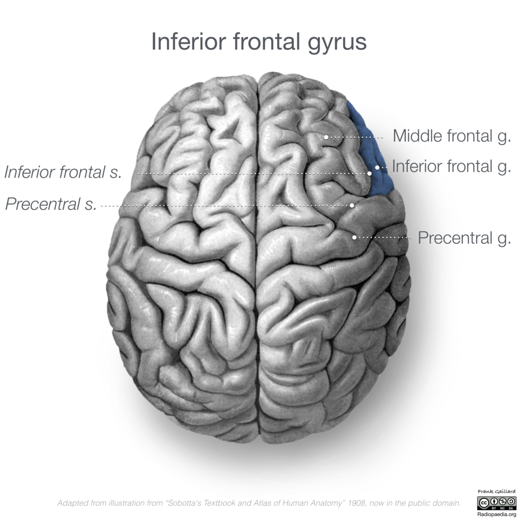
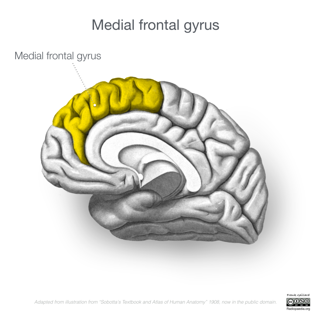
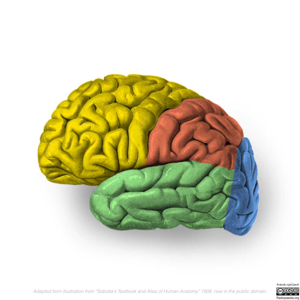
- The Prefrontal Cortex (PFC): Involved in complex cognitive behaviors, including decision making and moderating social behavior. The PFC plays a critical role in empathy, allowing us to understand others’ perspectives and feelings.
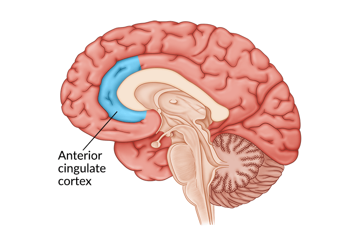
- The Anterior Cingulate Cortex (ACC): Associated with emotional processing, the ACC is activated when individuals experience personal distress or empathize with others’ pain.
- The Insula: This region is crucial for processing emotions and bodily awareness. It becomes activated when individuals feel compassion, facilitating a connection between the observation of suffering and the emotional response it elicits.
- The Amygdala: Known for its role in fear and emotion processing, the amygdala also contributes to identifying emotional states in others, which is essential for empathic concern and compassion.
The Role of Mirror Neurons
Another fascinating discovery in the neuroscience of compassion involves mirror neurons, which fire both when an individual acts and when they observe the same action performed by another. This mirroring mechanism is thought to underpin the capacity for empathy, enabling individuals to ‘feel into’ others’ experiences. fMRI studies examining the activity of mirror neurons during compassionate responses have provided insights into how we understand and share the emotions of others.
Compassion Training and Its Neural Correlates
Interestingly, research utilizing fMRI has also explored the impact of compassion training on brain function. Practices such as loving-kindness meditation (LKM) and compassion meditation have been shown to enhance compassionate feelings and altruistic behavior. fMRI studies conducted on individuals who engage in these practices regularly have observed significant changes in brain activity.
- Increased Activation in Compassion-Related Brain Areas: Individuals with extensive compassion meditation experience show increased activation in the insula, ACC, and PFC when exposed to sounds of distress or when practicing compassion meditation. This suggests that compassion training can enhance emotional resonance with others’ suffering and promote a more compassionate response.
- Structural Changes: Long-term practitioners of compassion meditation exhibit structural changes in brain regions associated with compassion, including increased gray matter density in the PFC and insula. These changes indicate that compassion training can lead to enduring alterations in the brain, potentially enhancing an individual’s capacity for empathy and compassion.
Compassion, Altruism, and the Social Brain


The investigation into the neural basis of compassion also sheds light on the broader concept of altruism – the selfless concern for the well-being of others. fMRI studies have demonstrated that acts of kindness and altruistic behavior activate reward centers in the brain, such as the ventral striatum, suggesting that helping others brings intrinsic satisfaction. This aligns with the notion that compassion and altruism are deeply rooted in our biology, serving as fundamental components of social bonding and cooperation.
Implications and Future Directions
The integration of fMRI research and compassion studies holds promise for various fields. In psychology and psychiatry, understanding the neural mechanisms of compassion can inform interventions for disorders characterized by deficits in social cognition and empathy, such as autism spectrum disorder and psychopathy. In medicine, compassion training for healthcare professionals could enhance patient care by fostering genuine empathic connections. Moreover, at a societal level, promoting compassion through education and policy could contribute to more cohesive and humane communities.
As we continue to unravel the neural underpinnings of compassion, future research will likely explore the genetic and environmental factors that influence these neural pathways. Additionally, the impact of virtual reality and other technologies on fostering empathy and compassion presents a novel area for investigation. By advancing our understanding of the brain’s role in compassion, we edge closer to harnessing its full potential for individual well-being and societal harmony.
In conclusion, the exploration of compassion through the lens of fMRI has illuminated the intricate neural networks that enable this pivotal human virtue. As we deepen our understanding of how the brain embodies compassion, we unlock new possibilities for nurturing empathy and altruism within ourselves and our communities, paving the way for a more compassionate world.